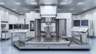
How to Choose the Right X Ray Intensifier for Optimal Imaging Performance
In the ever-evolving field of medical imaging, the choice of an X Ray Intensifier plays a crucial role in determining the quality of diagnostic images and, ultimately, patient care. According to a recent market analysis by Grand View Research, the global X-ray equipment market is projected to reach $13.1 billion by 2025, highlighting the growing importance of advanced imaging technologies. A well-selected X Ray Intensifier can significantly enhance image clarity, reduce radiation exposure, and improve operational efficiency, making it essential for healthcare providers to understand the various options available.
With numerous models and specifications on the market, this tutorial aims to guide you through the process of selecting the ideal X Ray Intensifier to achieve optimal imaging performance, ensuring that you make informed decisions that align with your clinical needs and technological advancements.
Key Factors to Consider When Selecting an X-Ray Intensifier
 When selecting an X-ray intensifier, several key factors play a crucial role in ensuring optimal imaging performance. Firstly, the material of the intensifier significantly impacts the quality of the images produced. Common materials include cesium iodide and columnar films, each offering different levels of resolution and light output. Choosing the right material will depend on the specific application, so it is essential to consider the imaging requirements to achieve the best results.
Another important aspect is the sensitivity and resolution of the intensifier. Higher sensitivity means better performance in low-dose situations, which is vital for patient safety and image quality. Additionally, the resolution determines how well the details of the image can be perceived. Evaluating the specifications provided by manufacturers can help identify the right balance between sensitivity and resolution tailored to your imaging needs. Ultimately, understanding these factors will guide you in choosing an X-ray intensifier that maximizes both performance and diagnostic effectiveness.
Understanding Different Types of X-Ray Intensifiers and Their Applications
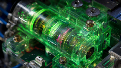
When selecting an X-ray intensifier, understanding the different types and their specific applications is crucial for achieving optimal imaging performance. There are primarily two types of X-ray intensifiers: cesium iodide (CsI) and sodium iodide (NaI). CsI is favored for its high conversion efficiency and reduced patient exposure due to its ability to provide clearer images with less radiation. This makes it ideal for diagnostic radiography and fluoroscopy, where image clarity is paramount.
On the other hand, NaI intensifiers are known for their superior light output and are often employed in specialized fields such as nuclear medicine. Their high sensitivity to X-rays allows for detailed imaging in low-dose scenarios, making them suitable for applications like gamma camera systems. Understanding these differences is essential for healthcare professionals seeking to optimize imaging technology in their practices, as the choice of intensifier can significantly affect the quality of the diagnostic output and patient safety.
How to Choose the Right X Ray Intensifier for Optimal Imaging Performance
| Type of X-Ray Intensifier |
Material |
Image Quality |
Application |
Advantages |
| Csl (Cesium Iodide) |
CsI(Tl) |
High |
Radiology, fluoroscopy |
Excellent spatial resolution |
| Gd2O2S (Gadolinium Oxysulphide) |
Gd2O2S:Tb |
Moderate |
Dental imaging, industrial radiography |
Lower cost, good light output |
| ZnS (Zinc Sulfide) |
ZnS:Ag |
Low |
High-energy applications |
Very low cost |
| Fiber Optic |
Glass fiber |
Variable |
Portable systems, veterinary imaging |
Flexibility and lightweight |
| Image Intensifier Tube |
Ceramic |
High |
C-arm systems, surgical imaging |
Compact size, high gain |
Evaluation of Imaging Performance: Sensitivity vs. Resolution
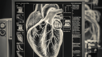
When evaluating X-ray intensifiers, the interplay between sensitivity and resolution is crucial for achieving optimal imaging performance. Sensitivity refers to the ability of the intensifier to detect low levels of X-ray radiation, which is essential for visualizing subtle details in images. A highly sensitive intensifier can produce clearer images from minimal exposure, thus reducing the risk of radiation damage to patients. This is especially important in fields such as oncology, where fine details can dictate the diagnosis and subsequent treatment plans.
On the other hand, resolution describes the intensifier's capability to display fine details and distinguish between closely spaced objects. High resolution is vital in scenarios where precise diagnosis is necessary, such as in the case of identifying early-stage diseases. However, there is often a trade-off between sensitivity and resolution; intensifiers that enhance one aspect may diminish the other. Therefore, choosing the right X-ray intensifier should focus on the specific imaging needs of the application, balancing these two performance metrics to ensure high-quality imaging that accurately supports clinical decisions.
How to Choose the Right X Ray Intensifier for Optimal Imaging Performance
This chart compares the sensitivity and resolution of different X-ray intensifiers, helping to evaluate their performance in imaging.
The Role of Material and Design in X-Ray Intensifier Efficiency
When selecting an X-ray intensifier, material and design play crucial roles in determining its efficiency and imaging performance. The choice of materials significantly affects the conversion efficiency of X-ray photons into visible light. For instance, traditional materials such as cesium iodide (CsI) have high attenuation properties, allowing for better image quality with less radiation exposure. According to a report by the Radiological Society of North America (RSNA), intensifiers using CsI can enhance image quality by up to 30% compared to those using older materials like calcium tungstate.
Design aspects, including the geometry and thickness of the phosphor layer, also influence the overall performance of X-ray intensifiers. Thinner layers can reduce scatter and improve resolution, while specific design configurations can optimize the light output for better detection sensitivity. A study published in the Journal of Medical Imaging highlighted that innovative designs could potentially increase the efficiency of X-ray intensifiers by over 50% in some applications, underscoring the importance of these factors in achieving optimal imaging performance. By considering both material properties and design innovations, healthcare professionals can significantly enhance the diagnostic capabilities of their imaging equipment.
Future Trends in X-Ray Intensifier Technology and Their Impact on Imaging
As the demand for high-quality imaging continues to rise, the future of X-ray intensifier technology is poised for significant advancements. The U.S. market for interventional X-ray and mobile C-arm equipment shows promising growth, recovering sharply in 2021 with projections indicating that it will exceed $4.9 billion by 2026. This surge highlights a shift towards more adept imaging solutions that meet stringent healthcare demands.
Tip 1: When selecting an X-ray intensifier, consider models that are designed for versatile applications. The evolving landscape suggests that the best systems integrate advanced features capable of diverse imaging requirements, enhancing both efficiency and efficacy.
Moreover, recent trends point towards innovations such as improved digital imaging software and enhanced sensitivity. These developments not only elevate image clarity but also reduce radiation exposure for patients. As such, selecting future-proof technology becomes paramount in ensuring optimal imaging performance.
Tip 2: Stay abreast of technological trends by engaging with industry reports and demonstrations. This knowledge can guide decision-making processes and ensure your investments align with future industry standards.