Selecting the best Mobile Image Intensifier in 2026 means looking beyond a product name or headline specification. In clinical imaging, the right mobile C-arm depends on the procedures performed, room layout, patient needs, and available support. A unit used for orthopedic imaging may face different demands from one used in pain management or operating rooms. Image clarity matters, but so do field of view, positioning flexibility, controls, and the time needed to move and prepare the system.
This guide compares practical factors that clinicians and procurement teams can verify: image performance, dose-management features, mobility, workflow, service availability, and total ownership costs. Specifications should be checked against manufacturer documentation and assessed by qualified imaging professionals. Where possible, demonstrations using realistic clinical tasks can reveal handling issues that a brochure will not. Small details count. A stiff control, awkward cable, or limited clearance can slow a busy room.
There is no single winner for every department. That answer can feel unsatisfying. Still, a careful comparison is more useful than declaring one model “best” without context. The discussion also recognizes that image intensifiers and newer detector designs may differ in performance, maintenance, and suitability. Buyers should confirm current configurations, local service arrangements, and compatibility before deciding. Some needs are hard to reduce to a score. The aim here is a clear, evidence-conscious starting point—not a substitute for clinical evaluation or a site-specific assessment.
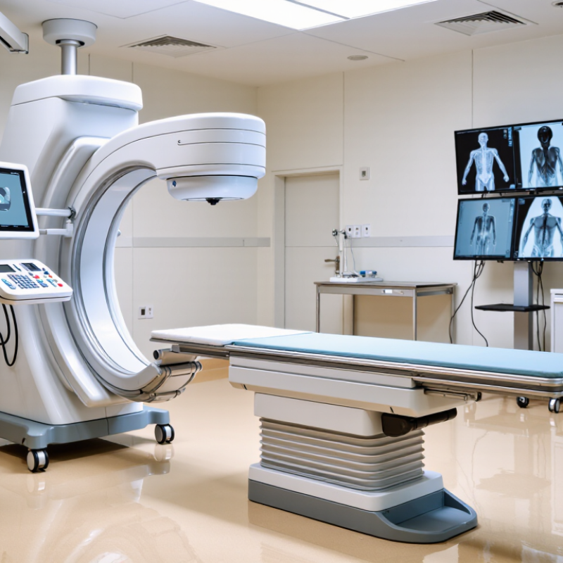
A mobile image intensifier is an X-ray imaging system on wheels, commonly used to guide procedures in operating rooms. It creates moving images rather than a single still picture. An X-ray tube sends radiation through the patient; tissues absorb different amounts, forming a pattern at the intensifier’s input screen. A photocathode converts that pattern into electrons. Electron optics focus and accelerate them toward an output screen, which produces a brighter image for a camera or display. The image can feel immediate. But it is not a direct view inside the body.
UNSCEAR’s 2020/2021 report estimates about 4.2 billion diagnostic radiological examinations worldwide over the period it assessed, underscoring the importance of imaging quality and radiation management. For a mobile system, image quality depends on more than brightness: positioning, field of view, movement, and exposure settings all matter. A technologist may adjust the unit around a crowded operating table, where even a small angle change can affect what appears on screen. Image intensifiers can also show edge distortion and reduced detail compared with newer detector designs. That trade-off deserves attention. In 2026, the “best” choice depends on the clinical task, image consistency, and how well staff can operate and maintain the system.
A mobile image intensifier should deliver usable detail at low dose, not merely a bright image.
Assess limiting resolution, low-contrast visibility, image uniformity, distortion, and field-of-view changes. Check performance with the unit positioned as it will be used: a slight angle or crowded bedside setup can expose weaknesses that a showroom image hides. AAPM Task Group 125’s report on fluoroscopic systems emphasizes evaluating image quality alongside automatic dose-rate control.
Dose matters. So does repeatability.
Portability counts, too. Look for smooth movement, reliable braking, quick positioning, and controls that remain clear under dim room lighting. The FDA notes that visible skin effects may occur around a peak skin dose of 2 Gy; this is a safety threshold, not a target. A unit’s dose display and protocols should help teams keep exposure appropriate for each examination.
Not just pixels.
One caveat: a sharper image may demand more radiation, so compare image quality at matched dose settings. In practice, the best choice is the system that gives consistent detail, fits real workflows, and makes dose trade-offs visible to staff. No single specification captures all three.
What Is the Best Mobile Image Intensifier in 2026?
Comparing mobile image intensifier models starts with the work they must support. Check image detail, field of view, and performance at different magnification levels. Ask for demonstrations using realistic clinical settings, not only polished sample images. Small details matter.
A useful comparison also includes dose controls, display readability, and how easily the unit moves around a crowded room. Check whether the C-arm clears the table and reaches the positions your team needs. Note startup time, battery runtime, and storage requirements. A technical specification can look impressive, while daily workflow tells a different story. Even a tidy scorecard can miss that gap.
Look closely at training, maintenance access, and service response times. Confirm that staff can understand dose information and use available dose-saving features correctly. Compare image quality under your facility’s established protocols, with qualified clinical and radiation-safety professionals involved. If possible, gather feedback from operators who will use the system regularly. Their comments may reveal awkward controls or limited visibility that a brief demonstration overlooks. No single model is best for every room; the right choice depends on clinical needs, workflow, support, and budget.
There is no single best image-intensifier size for every procedure. The comparison below uses common nominal input-field classes found in mobile fluoroscopy systems. These are general category descriptions, not specifications for individual products.
| Comparison Dimension | Compact 9-Inch Class | Standard 12-Inch Class | Large 16-Inch Class |
|---|---|---|---|
| Nominal input-field diameter | About 9 in (23 cm) | About 12 in (31 cm) | About 16 in (41 cm) |
| Field coverage | Smaller overall field; useful when the anatomy and working area are relatively limited. | Middle-ground coverage for a broad range of routine imaging tasks. | Larger overall field, which can help when a wider anatomical area must be viewed. |
| Typical workflow fit | Procedures where compact equipment and a smaller field are priorities, subject to clinical requirements. | General-purpose mobile fluoroscopy where a balance of coverage and maneuverability is needed. | Procedures that benefit from wider coverage, provided the room and equipment can accommodate the system. |
| Magnification modes | Check the number and size of selectable fields; magnification generally reduces the displayed field of view. | Check available field selections and whether they meet the intended anatomical and procedural needs. | Confirm that magnification options provide useful detail without sacrificing needed field coverage. |
| Image-intensifier technology | Uses an image-intensifier tube with an optical/electronic camera chain; image characteristics depend on the complete system. | Uses the same general image-intensifier architecture; compare the full imaging chain rather than size alone. | Uses the same general image-intensifier architecture; a larger input field does not by itself guarantee better image quality. |
| Image quality checks | Review measured spatial resolution, contrast performance, distortion, and image uniformity for the specific system. | Review the same acceptance-test results and compare them under equivalent imaging conditions. | Review the same acceptance-test results; assess image quality across the field, not only at the center. |
| Dose-management checks | Confirm available dose settings, pulsed-fluoroscopy options, last-image hold, and displayed dose information. | Compare dose-control functions and test performance at clinically relevant settings. | Compare dose-control functions carefully; field size alone does not determine patient dose. |
| Room and positioning considerations | Assess C-arm movement, clearance, working distance, and access around the patient and staff. | Check orbital movement, source-to-image distance, positioning range, and access to the operating area. | Pay particular attention to clearance, maneuverability, storage space, and access in the intended room. |
| What to verify before purchase | Actual selectable field sizes, measured image performance, dose documentation, service support, and compatibility with the clinical workflow. | Actual field sizes, image and dose test results, positioning range, software functions, and total cost of ownership. | Actual field sizes, room fit, positioning performance, image and dose test results, and installation requirements. |
| Best fit when… | A smaller field meets the clinical task and compact operation is a priority. | A versatile balance of field coverage and day-to-day usability is preferred. | Wider field coverage is important and room layout and system handling remain practical. |
How to choose: Compare systems using the same test conditions and intended clinical tasks. Confirm the manufacturer’s stated field sizes, acceptance-test measurements, dose-control features, room requirements, and applicable regulatory documentation. Nominal input-field size is not the same as the usable image field, and system performance varies by configuration.
The best mobile image intensifier depends on the room, procedure, and staff workflow. For orthopedic cases and small operating rooms, a compact C-arm with easy positioning can reduce awkward repositioning around the table. For abdominal imaging, a larger field of view may be more useful. In pain clinics, smooth movement and clear low-dose settings can matter more than maximum output. Small details count: Can the monitor be seen over a drape? Does the unit fit beside anesthesia equipment?
UNSCEAR’s 2020/2021 report estimates about 4.2 billion diagnostic radiological examinations worldwide each year. That scale makes dose awareness and consistent quality checks practical buying criteria, not optional extras. AAPM Report No. 125 also describes quality control practices for diagnostic radiology equipment. Compare image quality at the settings your team actually uses, and ask how service checks are documented. I would not choose from a brochure alone; room layout and staff experience can change the answer.
Tips: Test the unit in your real procedure room. Check maneuverability, monitor visibility, pulse settings, and image clarity on a typical patient. Then review maintenance and dose records with a medical physicist.
A mobile image intensifier should fit the procedure, room, and clinical workload—not just promise a sharp image. Check image quality across the field of view, especially at the edges, where distortion can affect positioning. Ask for measured performance on a test phantom, not a showroom demonstration. AAPM Report No. 125 describes image-quality and radiation-output checks as part of diagnostic imaging quality control. That makes documented acceptance and routine testing practical selection criteria.
Compare field-of-view options, pulsed fluoroscopy settings, dose displays, and how easily staff can position the detector around a patient. Review the service plan, too: downtime matters when one unit serves several rooms. The FDA’s fluoroscopy performance standard specifies an audible cumulative-timer signal at five-minute intervals. Treat this as one safety feature, not proof of low dose. A brighter image can still come with avoidable exposure. It is easy to overvalue a crisp demo; ask for dose and image-quality results under realistic operating conditions.
Tips: Test the unit in a room with your usual table and accessories. Record dose-area product and image quality at typical settings, then compare systems on the same phantom. Check whether controls remain reachable while positioning the unit. Small workflow annoyances add up. A convenient feature may matter less than dependable service—worth questioning before purchase.
Reading the chart: These nominal 6-, 9-, and 12-inch input-field formats are shown as centimeter conversions, not as a ranking of systems. A larger field covers more anatomy at once; a smaller field or magnification mode may be useful when more detail is needed. When comparing mobile systems, also assess image quality, dose management, maneuverability, display usability, and service requirements.