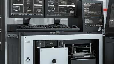
Choosing a C Arm Intensifier in 2026 requires more than comparing detector size or purchase price. It requires clinical judgment, radiation awareness, and a realistic service plan. The global C-arm market is expanding, although estimates differ by research firm. Grand View Research projects continued growth through 2030, driven by orthopedic, cardiovascular, pain-management, and trauma procedures. These figures show demand. They do not guarantee a suitable device for every hospital.
Technical details matter at the operating table. Consider image quality at low dose, pulse rates, field of view, thermal capacity, mobility, and compatibility with surgical tables. A compact unit may fit a crowded theater better. A larger detector may reduce repositioning during complex procedures. Check the monitor brightness under surgical lights. Test the wheels on uneven flooring. Small details become expensive problems.
Radiation protection must remain central. The International Atomic Energy Agency emphasizes optimization and the ALARA principle in fluoroscopy. As radiation-protection specialist Dr. Madan M. Rehani has stated, “Radiation protection is not about avoiding radiation; it is about using it wisely.” That principle applies directly to C-arm selection. Ask vendors for dose-area-product displays, dose alerts, staff training, and service response times. A system with impressive specifications may still perform poorly without skilled users. This is where buyers often underestimate risk. No product comparison is perfect. Procurement teams should review clinical workflow, total ownership cost, cybersecurity, training, and local regulatory requirements before signing. The best C Arm Intensifier is not always the most advanced model. It is the one that delivers dependable imaging, controlled exposure, and practical support every day.
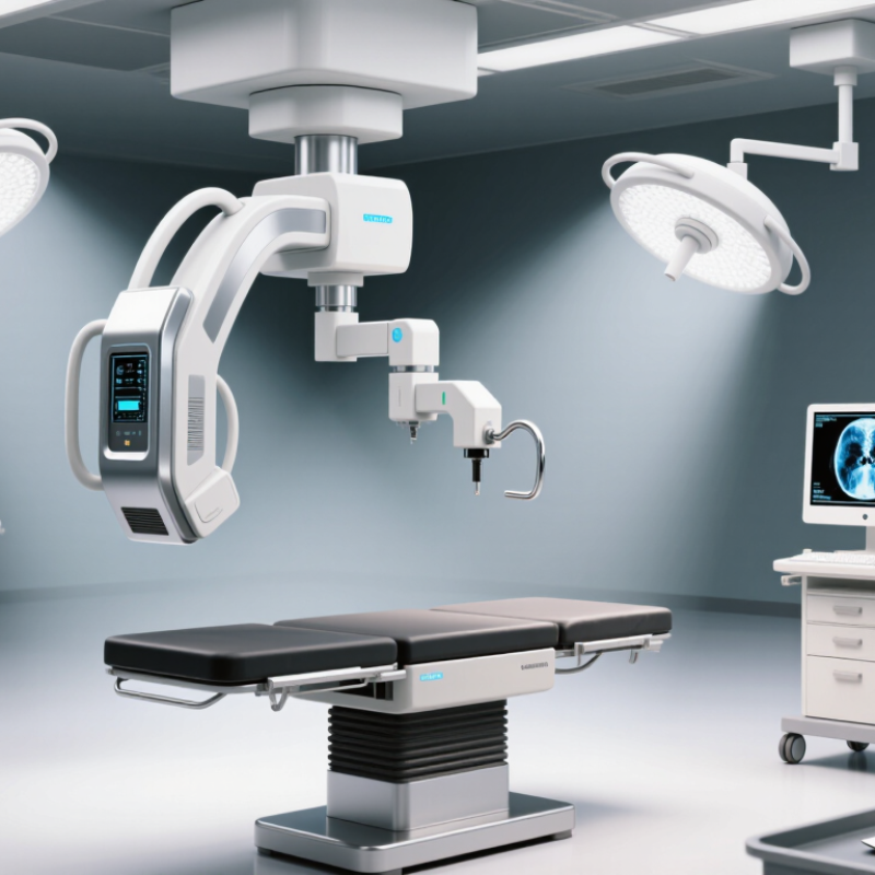
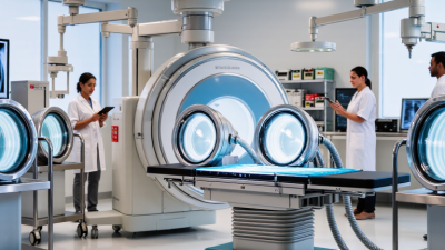
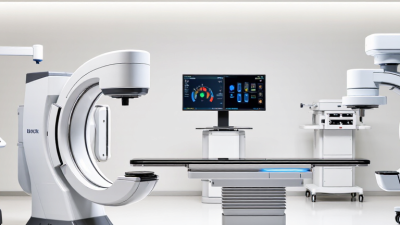
A C-arm intensifier converts X-rays passing through the patient into a visible image for the clinical team. In an image-intensifier system, the detector amplifies that image before it appears on the monitor. The C-shaped arm lets staff position the X-ray source and detector around the patient. Small movements matter. A few centimeters can change the view of a joint, implant, or instrument.
The intensifier affects image brightness, detail, and usable field of view, but it does not work alone. Image quality also depends on patient size, positioning, exposure settings, and display calibration. The FDA describes fluoroscopy as real-time X-ray imaging and advises using the lowest exposure needed for the task. That guidance is practical at the bedside. NCRP Report No. 160 estimated that medical procedures contributed about 48% of the average U.S. population’s radiation exposure in 2006; this figure covers medical exposure broadly, not C-arm use alone. When comparing systems, review image uniformity, distortion near the edges, field-of-view options, and service history. Ask to inspect images at the edges, not just the center. Specifications can look reassuring. Real clinical images may tell a less tidy story.
Choosing a C-arm intensifier in 2026 starts with the work it must support. An orthopedic team checking screw placement needs clear views of bone and hardware, often from several angles. Pain-management procedures may require precise visualization of needles and contrast, with tight control of radiation exposure. Vascular tasks can demand different contrast performance and workflow. A single specification sheet rarely captures these differences. Ask clinicians to describe typical cases, patient sizes, required projections, and room constraints. Then test representative images in the actual clinical environment.
Radiation protection belongs in that assessment. The International Atomic Energy Agency notes that prolonged fluoroscopic procedures can produce skin doses associated with tissue reactions; effects may occur around 2 Gy, depending on dose distribution and patient factors. That is not a universal safety cutoff. It is a reason to examine dose displays, pulsed fluoroscopy, collimation, and operator training. The FDA also advises facilities to use the lowest exposure needed for adequate imaging. Compare systems using realistic tasks, not just maximum output or a showroom image. Check whether staff can position the unit without blocking access, and whether images remain useful at the needed distance. One awkward detail: teams may disagree about what “good image quality” means. Record those disagreements, review sample cases, and revisit the choice after clinical use.
Image quality should be judged on anatomy, not showroom brightness. Check low-contrast visibility, spatial resolution, lag, and distortion with a realistic phantom. The IAEA recommends acceptance testing and routine quality control because detector performance can drift over time. Ask for measured results at common surgical settings. Numbers without test conditions can mislead.
Field of view changes the clinical picture. A larger field captures more anatomy, which helps during positioning and complex procedures. A smaller field improves detail but may require repositioning. Magnification can also increase patient exposure. The IAEA’s Radiation Protection of Patients guidance links tighter collimation with better dose efficiency. Test a 9-inch and 12-inch mode on the same phantom. The difference becomes obvious.
Dose controls deserve daily attention. Look for automatic brightness control, pulsed fluoroscopy, adjustable pulse rates, last-image hold, and clear cumulative dose displays. ICRP Publication 85 associates temporary skin effects with doses near 2 Gy and more serious injury at higher levels. These are not remote numbers during lengthy interventions. The FDA also advises monitoring dose and recording exposure indicators when appropriate. Still, automatic control is not magic. It may raise output when the table, patient size, or projection changes. In my view, the best system is the one staff can understand quickly, even under pressure. Some specifications remain surprisingly vague. Ask for independent testing data.
Choosing a C-arm intensifier in 2026 starts with the room, not the brochure. Measure doorway widths, ceiling height, and available power connections before comparing specifications. Confirm compatibility with the C-arm generator, mounting system, image-processing software, and surgical table. A mismatched interface can create delays, even when the image quality looks impressive. Ask for demonstrations using realistic anatomy and typical operating-room positions. Watch how quickly staff can move, lock, expose, and review images. Small controls matter during a crowded procedure. They matter more under pressure.
Workflow should remain central to the evaluation. Check whether the intensifier supports your current monitors, data formats, and secure network practices. Confirm dose display, image storage, and export functions with your clinical and technical teams.
Service support deserves the same attention as hardware. Request written response times, preventive maintenance schedules, spare-part availability, and local technician qualifications. Ask how software updates are tested before installation.
Talk with users who have operated the system for several months, not only during a sales demonstration. My own checklist has missed cable routing before. That mistake taught me to inspect the complete setup in the actual room. No system is perfect. Reliable support can make an imperfect system workable.
Safety standards, budget, and future requirements should shape a C-arm intensifier purchase. Ask for current documentation on radiation protection, electrical safety, and image performance. Requirements vary by region and facility, so confirm them with your medical physicist or qualified compliance lead. Check the unit’s shielding, dose display, emergency controls, and service records. Ask how acceptance testing will be performed after installation.
Look beyond the purchase price. Include installation, staff training, maintenance, replacement parts, and possible room modifications in the budget. Request a written service plan and realistic repair timelines. A low-cost system can become expensive when a key component is unavailable. It happens.
Future needs matter, too. Compare detector options, image-processing features, software support, and compatibility with your existing workflow. Consider the procedures you expect to perform, not just today’s case mix. Ask whether upgrades are practical, and how long security and software updates are expected. A larger display may seem minor, but it can improve visibility during long procedures. Still, more features are not always better. I would test image quality and controls with the staff who will use the system, under realistic room conditions, before committing. A short demonstration may not reveal every limitation.