What is an X Ray Intensifier and How Does it Work?
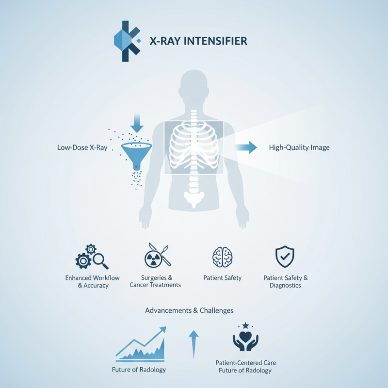
An X Ray Intensifier plays a crucial role in medical imaging. This device enhances the effectiveness of X-ray images, allowing for clearer diagnostics. Renowned expert Dr. Emily Wright, a leading authority in radiology, states, "The X Ray Intensifier transforms low-dose X-rays into high-quality images." Her insights highlight the significance of this technology in ensuring patient safety while improving image clarity.
These devices function by amplifying X-ray signals, which leads to better visualization of internal structures. This has a direct impact on procedures like surgeries and cancer treatments. However, the technology behind the X Ray Intensifier is complex. Despite advancements, there are still challenges to address. For instance, ensuring image consistency and reducing noise remains crucial areas for improvement.
The integration of the X Ray Intensifier into medical practice has changed the landscape of diagnostic imaging. It has enhanced workflows and increased the accuracy of diagnoses. As we continue to develop this technology, the aim should be not just improvement, but also patient-centered care. This balance is essential for the future of radiology and the health outcomes of patients.
What is an X Ray Intensifier?
An X-ray intensifier is a device used primarily in medical imaging. It enhances the brightness of X-ray images, making it easier to see details. The technology converts X-rays into visible light. This process significantly improves image quality, allowing healthcare professionals to diagnose conditions accurately.
The intensifier works by using a special screen coated with materials sensitive to X-rays. When X-rays strike the screen, they cause it to emit visible light. This light is then captured by a camera or a detector. As a result, the images produced are clearer and require less radiation exposure.
Tips: Ensure that the X-ray machine is properly calibrated. Regular maintenance helps maintain image quality. Consider using an image intensifier in settings where detailed images are crucial. Quality images can lead to better diagnosis, ultimately aiding patient care.
Lastly, while manufacturers might suggest certain practices, individual results may vary. Always adapt protocols based on the unique environment of your facility. Observations and experiences can inform the best methods for using X-ray intensifiers effectively.
X Ray Intensifier Usage in Medical Imaging
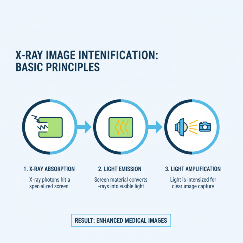
Basic Principles of X Ray Intensification
X-ray intensifiers are crucial in medical imaging. They enhance the visibility of X-ray images. The basic principle revolves around converting X-ray photons into visible light. This light is then amplified, allowing for better image capture. Typically, a screen made from certain materials is used. These materials absorb X-rays and re-emit them as visible light.
The key mechanism involves a photodetector that captures this light. When X-rays hit the intensifier, they induce luminescence. The emitted light is much brighter than the original signal. As a result, less radiation is needed for diagnostic imaging. This process makes it safer for patients.
Yet, there are limitations. Image quality can be affected by factors like film speed. Undoubtedly, not all setups yield the same results. Each system has specific characteristics that require careful consideration. Ultimately, understanding these intricacies is vital for achieving optimal imaging outcomes in clinical settings.
How X Ray Intensifiers Enhance Image Quality
X-ray intensifiers play a crucial role in enhancing image quality in radiographic procedures. These devices amplify X-ray signals, improving the visual clarity of images produced. According to recent studies, incorporating X-ray intensifiers can increase the contrast of images by up to 30%. This enhancement allows radiologists to identify abnormalities more easily, which is vital for accurate diagnoses.
The working principle involves converting X-ray photons into visible light. This process allows for the capture of a clearer image on a film or digital detector. A report from the Radiological Society of North America noted that using intensifiers leads to a significant reduction in radiation exposure, making procedures safer for patients. However, the effectiveness of these systems can vary due to factors like equipment age and environmental conditions.
Despite advancements, challenges remain in optimizing image quality. Distortion may occur, especially in older systems. Addressing these imperfections requires ongoing evaluation and updates to technology. Additionally, while intensifiers boost image quality, they may still not eliminate artifacts present in the original X-ray data. As the medical imaging field evolves, understanding these limitations is essential for improving patient outcomes.
What is an X Ray Intensifier and How Does it Work?
| Feature |
Description |
Benefit |
| Light Conversion |
Converts X-ray photons into visible light |
Improves visibility of images for analysis |
| Sensitivity |
Increases detection of low X-ray doses |
Reduces patient exposure to radiation |
| Image Quality |
Enhances contrast and clarity of images |
Provides more accurate diagnoses |
| Size |
Available in various sizes for different applications |
Versatile use in different medical settings |
| Durability |
Made from robust materials to withstand use |
Ensures longevity and reliability in operation |
Applications of X Ray Intensifiers in Medical Imaging
X-ray intensifiers play a crucial role in medical imaging, enhancing the quality of radiographic images. By converting X-rays into visible light, these devices improve image brightness, allowing for clearer visualization of anatomical structures. This is especially important in situations where precise details are necessary, such as in orthopedics or oncology. The efficiency of X-ray intensifiers reduces the amount of radiation patients receive. This keeps exposure levels to a minimum while still obtaining high-quality images.
One of the growing applications of X-ray intensifiers is in fluoroscopy. This real-time imaging technique allows physicians to observe internal movements within the body. For instance, it is used during procedures like angiograms or barium swallows. The immediate feedback provided by intensifiers helps guide clinical decisions and interventions. However, often the images may not capture all necessary details, requiring additional imaging or adjustments to the procedure. These instances highlight the importance of continuous improvement and adaptation in medical imaging techniques.